
Among the many most used diagnostic assessments, the magnetic resonance imaging (MRI) permits to acquire detailed photos in excessive decision of the within of our physique, by means of the usage of magnetic fields with out ionizing radiation. Because of its precision, it’s now utilized in quite a few fields, to detect varied pathologies and research their course.
On this article we are going to attempt to perceive the way it works, during which instances it’s helpful for analysis and if there are any contraindications in its use.
MRI: how does this examination work?
The magnetic resonance investigation technique, found in 1946, was first utilized within the medical subject in the course of the Nineteen Seventies by Raymond Vahan Damadian. The physician sensed that the usage of this method might be notably efficient within the research of most cancers, hypothesizing that tumors and tissues may give totally different responses to MRI.
Primarily based on the “physics of magnetic fields”, the operation of the magnetic resonance gear is quite complicated, for non-specialists.
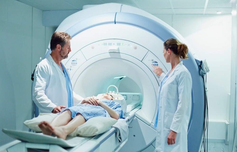
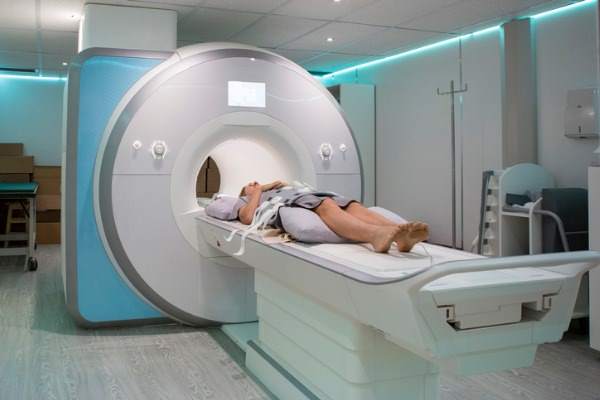
First, to carry out the examination, the affected person should lie down on a mattress, which is slid inside a cylindrical equipment, and stay relaxed and motionless during the investigation.
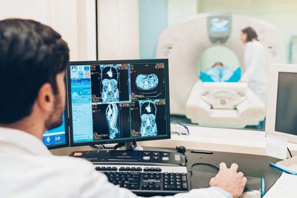
The equipment makes use of magnetic fields intense together with radio frequency pulses. Electromagnetic waves quickly alter the atoms of which the tissues of our organs are composed, which purchase power and orient themselves in line with the pattern of the magnetic subject. When the latter is deactivated, the atoms return to their regular orientation, giving up power and emitting a sign. It’s exactly this sign that’s picked up by the gear, processed and eventually changed into Pictures.
The machine consists of a hole cylinder with magnets inside that are used to generate beams of radio waves helpful for examination. The basic side for the success of the MRI is that the affected person stays nonetheless for your complete length of the investigation, which is determined by the extent of the a part of the physique to be examined and might be as much as 45 minutes.
MRI means that you can have one three-dimensional view of the physique, since, by exploiting biochemical processes, sections of the physique are obtained not solely within the axial aircraft, but in addition within the frontal, lateral, indirect aircraft and from totally different angles.
Distinction MRI
MRI can also be prescribed with the usage of a distinction medium, that’s, of a paramagnetic substance (resembling gadolinium) which is injected right into a vein, to be able to extra precisely detect neoplastic and inflammatory pathologies. Any such magnetic resonance, in comparison with the standard one, is ready to present clearer and extra detailed photos of the inner constructions of our physique. Because of this, it’s used to check tumors, the blood stream within the arteries and veins, the blood provide of an organ and accidents to inner anatomical constructions in higher depth.
However how does it work? The distinction brokers are distributed within the vessels and organs: gadolinium molecules alter the molecular properties of the magnetic subject of the constructions during which they’re situated, modifying the sign and picture of the magnetic resonance. On this approach, the vessels and eventual organ lesions are visualized, accentuating the variations within the tissues and vascularity.
When is MRI prescribed?
Magnetic resonance imaging is now used to acquire detailed, high-resolution photos of quite a few tissues. It thus proves to be a very helpful process in a number of fields, since technological advances through the years have made it one of many principal diagnostic imaging strategies.
Magnetic resonance imaging in neurology and neurosurgery
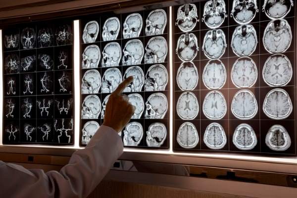
MRI supplies higher decision of nerve constructions than CT, leading to important visualization cranial nerves, mind stem harm And spinal wire. It’s also notably helpful for figuring out spinal alterations (tumors, abscesses) that compress the spinal wire and require pressing intervention. As well as, it’s indicated for the detection of demyelinating plaques, early coronary heart assaults, subclinical cerebral edema and cerebral contusions.
Magnetic resonance imaging in traumatology and orthopedics
Utilized in joint well being evaluation, joint MRI can be utilized for in-depth research of: elbow, shoulder, wrist, hand, knee, ankle and foot. It means that you can get detailed photos of the bone, cartilage, ligament and muscle element joints, thus offering data on traumatic, inflammatory and degenerative pathologies.
Magnetic resonance imaging in oncology
MRI is used for analysis and analysis of Stadium and of response to remedy of various kinds of tumor, permitting to hint an outline not solely morphological, but in addition structural of the tissues. It identifies the tumor not solely as a overseas “mass” throughout the physique, but in addition as an space of altered signaling within the context of an organ or tissue, with out essentially altering its quantity or profile of the identical.
Magnetic resonance imaging in cardiology
Additionally utilized within the cardiology subject, magnetic resonance imaging can be utilized for the analysis of pathologies affecting the coronary heart and of coronary heart valves resembling: ischemic coronary heart illness, dilated cardiomyopathies, myocarditis, hypertrophic cardiomyopathies, congenital coronary heart illness, valvular illness and pericardial illness. MRI of the center, by means of the usage of distinction, is the one investigation that right this moment permits us to view the presence of structural harm to this organ, each earlier and up to date: a coronary heart assault, a myocarditis, irritation of the pericardium, and many others.
Magnetic resonance imaging in gastroenterology
Helpful within the analysis of illnesses of thestomach and of pelvis (liver and uterus), MRI can be used to visualise and research thesmall gut. This examination permits, in reality, to guage the doable presence of intestinal pathologies, whether or not it’s inflammatory or tumor-related illnesses.
It’s the reference approach for the analysis of continual inflammatory illnesses resembling Crohn’s illness and ulcerative colitis, however additionally it is helpful in evaluating any pathologies of an infectious nature resembling gastroenteritis and the presence of any tumors.
MRI: danger components and contraindications
Though the MRI represents a very innocent diagnostic investigation for our well being, it’s essential to know that not everybody can bear one of these investigation. There are in reality various kinds of contraindications.
Absolutely the contraindications
Sufferers present process surgical procedure, the place they had been used vascular clips iron-magnetic intracranial on aneurysms, can’t bear magnetic resonance imaging. The magnetic subject generated, in reality, may have a mechanical impact on the clips, inflicting a cerebral hemorrhage.
Moreover, this investigation can be not indicated in sufferers with:
- cardiac pacemakers
- coronary heart valves
- steel constructions resembling prostheses and screws
In all these instances, in reality, the examination will not be carried out to forestall the magnetic subject produced by the machine from shifting these constructions to a different location.
Sufferers who’ve had an accident previously or have carried out specific work actions with out safety (resembling within the metalworking sector) it’s at all times advisable to take an x-ray earlier than present process an MRI scan, to be able to exclude the presence of steel our bodies in your physique.
It’s also vital to contemplate what might be outlined as relative contraindications: particular instances, during which the benefits and drawbacks of one of these diagnostic examination have to be balanced. A affected person with a one magnetic sphincter who needs to bear an MRI scan needs to be conscious that this intracorporeal system might inactivate because of the investigation. For a similar purpose, a affected person utilizing the intrauterine spiral as a contraceptive technique, after having achieved the examination, you should go to the gynecologist to test that the system is within the appropriate place.
Now that we’ve seen how magnetic resonance imaging represents a elementary examination for the analysis and analysis of quite a few pathologies, all that continues to be is to decide on a facility with a staff of docs and specialised operators to carry out this necessary diagnostic investigation. Exactly for that reason, UniSalute affords 5 Particular person insurance coverage, to answer the totally different wants of everybody within the well being sector. Do you already know them?
Sources:
materdomini.it
Fondazioneveronesi.it
airc.it
msdmanuals.com
sandonato.grupposandonato.it


